Abstract:
Background: Cerebral small vessel disease (CSVD) is one of the most common forms of cerebrovascular disease in older adults and a frequent comorbidity in patients who have had COVID-19. SARS-CoV-2 infection can aggravate cerebral small-vessel damage through endothelial dysfunction, hypercoagulability and systemic inflammation, which may accelerate cognitive decline. Quantitative electroencephalography (qEEG) is an inexpensive, non-invasive and widely available tool for detecting functional brain changes, but its value in post-COVID CSVD has not been sufficiently studied.
Objective: To characterize qEEG changes in patients with CSVD who had recovered from COVID-19 and to assess their relationship with cognitive impairment and the severity of the past infection.
Methods: A total of 120 patients with CSVD (mean age 64.7 ± 7.2 years) examined at NEVROMED-SERVIS clinic (Namangan, Uzbekistan) were enrolled. The main group included 70 patients with laboratory-confirmed past COVID-19 (PCR, serology or medical records); the comparison group included 50 patients with CSVD and no history of COVID-19. CSVD was diagnosed on the basis of clinical, MRI and vascular criteria. Resting-state EEG (19 channels, 10–20 system) was recorded and analyzed quantitatively: peak alpha frequency (PAF) in O1/O2, alpha index, the (delta + theta)/(alpha + beta) ratio (DTABR) by region, the pairwise-derived brain symmetry index (pdBSI) and interhemispheric theta/alpha coherence (F3–F4, T3–T4, P3–P4, O1–O2). Cognition was assessed with the Montreal Cognitive Assessment (MoCA) and fatigue with the MFI-20 scale.
Results: Compared with the comparison group, post-COVID patients had a lower PAF (8.6 ± 0.7 vs 9.3 ± 0.6 Hz, p < 0.001), a lower alpha index (42% vs 54%, p < 0.001) and a higher frontal DTABR (2.14 ± 0.61 vs 1.62 ± 0.48, p < 0.001). Diffuse EEG slowing was found in 61% vs 34% of patients, respectively. pdBSI was higher (0.112 vs 0.081, p < 0.01), and frontal interhemispheric alpha coherence was reduced, indicating disrupted functional connectivity. Mean MoCA score was lower in the main group (21.4 ± 3.1 vs 23.8 ± 2.7, p < 0.001). PAF correlated positively (r = 0.52) and frontal DTABR negatively (r = −0.47) with MoCA scores. The most pronounced qEEG changes were observed in patients who had moderate-to-severe COVID-19.
Conclusions: Past COVID-19 in patients with CSVD is associated with slowing of background brain activity, increased interhemispheric asymmetry and reduced functional connectivity, and these changes are related to cognitive impairment. PAF and frontal DTABR may serve as simple, objective qEEG markers for identifying post-COVID patients with CSVD at higher risk of cognitive decline and for monitoring the effect of treatment and rehabilitation.
Keywords: Cerebral small vessel disease, COVID-19, quantitative EEG, peak alpha frequency, cognitive impairment.
Biography:
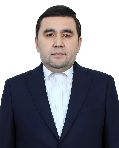
Dr. Ulugbek G. Askhonov is a neurologist with about 20 years of clinical experience in pediatric and adult neurology. He graduated with honors from Andijan State Medical Institute and completed his residency in pediatric neurology at the Tashkent Institute of Postgraduate Medical Education. He works as a neurologist at NEVROMED-SERVIS clinic. He is currently a PhD candidate studying the clinical and neurological features of Cerebral small vessel disease in patients after COVID-19.
© 2026 Mathews International LLC. All rights reserved.
